Ultrasound is one of the most commonly used imaging techniques in gynecology. It helps doctors assess various conditions affecting the uterus, including fibroids and cancer. Many women diagnosed with fibroids wonder if ultrasound can reliably distinguish these noncancerous growths from malignant tumors. This article explores the capabilities and limitations of ultrasound in identifying the differences between fibroids and cancer.
What Are Fibroids?
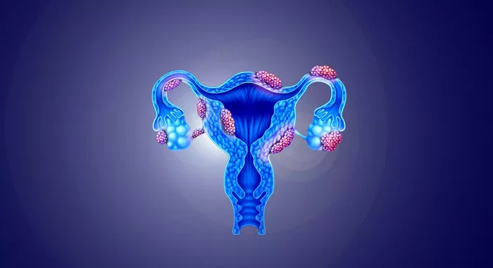
Fibroids are benign (noncancerous) tumors that grow in or around the uterus. They are made of muscle and fibrous tissue and can develop in different areas of the uterine wall. Fibroids vary in size, ranging from tiny, undetectable growths to large masses that distort the shape of the uterus.
Fibroids are very common in women of reproductive age. Many women have fibroids without experiencing any symptoms. However, when symptoms occur, they may include heavy menstrual bleeding, pelvic pain, frequent urination, and discomfort during sex.
What Is Uterine Cancer?
Uterine cancer refers to malignancies that develop in the uterus. The most common type is endometrial cancer, which starts in the lining of the uterus. Another type, uterine sarcoma, arises from the muscle or connective tissues of the uterus. Uterine cancer is less common than fibroids, but it is a serious condition that requires early detection and treatment.
Symptoms of uterine cancer may include abnormal vaginal bleeding, postmenopausal bleeding, pelvic pain, and unexplained weight loss. Since fibroids and uterine cancer can both cause irregular bleeding and pelvic pain, distinguishing between them is crucial.
How Does Ultrasound Work?
Ultrasound uses high-frequency sound waves to create images of internal organs. In gynecology, two main types of ultrasound are used:
Transabdominal Ultrasound
A transabdominal ultrasound involves placing a probe on the abdomen to capture images of the uterus and other pelvic structures. This method provides a broad view of the reproductive organs but may not always detect small abnormalities clearly.
Transvaginal Ultrasound
A transvaginal ultrasound involves inserting a probe into the vagina for a closer and more detailed view of the uterus. This technique is often preferred when evaluating fibroids, endometrial thickening, or suspicious growths.
Both types of ultrasound help doctors visualize the size, shape, and texture of uterine structures. However, determining whether a growth is benign or malignant based solely on ultrasound can be challenging.
Can an Ultrasound Differentiate Fibroids from Cancer?
Ultrasound can provide important clues about whether a uterine mass is likely to be a fibroid or cancerous tumor. However, it is not always definitive. Certain ultrasound features can suggest a higher likelihood of malignancy, while others indicate a benign fibroid.
Typical Ultrasound Appearance of Fibroids
Fibroids usually appear as well-defined, round, or oval masses within the uterus. They have a uniform, solid texture and may contain areas of calcification (hard deposits). Fibroids often cause the uterus to enlarge but maintain a smooth outer shape.
Typical Ultrasound Appearance of Uterine Cancer
Malignant tumors tend to have irregular shapes and poorly defined borders. They may appear more heterogeneous, meaning they have mixed solid and cystic (fluid-filled) components. Uterine cancers, especially sarcomas, may invade nearby tissues rather than staying contained within the uterus.
While these characteristics can help guide a diagnosis, they are not always definitive. Some fibroids can undergo degeneration, leading to irregular appearances on ultrasound, which may resemble a cancerous mass.
Limitations of Ultrasound in Diagnosing Uterine Cancer
Ultrasound is a valuable tool, but it has limitations when distinguishing fibroids from cancer. In some cases, additional testing is required to confirm a diagnosis.
Overlapping Features
Some fibroids can have atypical appearances, making them look similar to cancerous growths. Likewise, some early-stage cancers may resemble benign fibroids.
Inability to Confirm Malignancy
Ultrasound can suggest that a mass has concerning features, but it cannot definitively confirm whether a tumor is cancerous. A biopsy or further imaging may be needed.
Dependence on Operator Skill
The accuracy of an ultrasound depends on the expertise of the technician or radiologist interpreting the images. Misinterpretation can lead to unnecessary worry or missed diagnoses.
When Additional Testing Is Needed
If an ultrasound reveals a suspicious mass, further tests may be required to determine if the growth is cancerous.
MRI (Magnetic Resonance Imaging)
MRI provides a more detailed view of uterine structures than ultrasound. It can help distinguish fibroids from cancer by analyzing tissue composition and vascular patterns.
Endometrial Biopsy
If abnormal thickening of the uterine lining is detected, an endometrial biopsy may be performed. This involves collecting a small tissue sample from the uterine lining to check for cancer cells.
Hysteroscopy
A hysteroscopy allows direct visualization of the uterine cavity. A small camera is inserted through the cervix to examine any abnormal growths, and tissue samples can be taken for biopsy.
Doppler Ultrasound
Doppler ultrasound evaluates blood flow within a mass. Cancerous tumors tend to have irregular and increased blood supply, while fibroids typically have more organized blood flow.
What Should You Do If You Have a Uterine Mass?
If an ultrasound detects a mass in your uterus, your doctor will assess its characteristics and recommend further evaluation if necessary. It is important to follow up on any abnormal findings, especially if you experience unusual symptoms such as postmenopausal bleeding or persistent pelvic pain.
Monitoring Fibroids
If the ultrasound suggests a benign fibroid and there are no severe symptoms, monitoring may be recommended. Regular follow-ups ensure that the fibroid does not grow rapidly or change in appearance.
Seeking Further Testing
If the ultrasound raises concerns about possible malignancy, additional imaging or biopsy may be required to confirm the diagnosis. Early detection of uterine cancer significantly improves treatment outcomes.
Conclusion
Ultrasound is a useful tool for identifying uterine masses, but it has limitations in definitively distinguishing fibroids from cancer. While certain ultrasound features suggest whether a mass is benign or malignant, additional testing may be necessary for an accurate diagnosis. Women experiencing abnormal symptoms should consult their healthcare provider for proper evaluation and management.
Related topics:


